Full Content

ECTRIMS 2018 have been an opportunity to understand better the clinical expert needs

European Committee for treatment and research in Multiple Sclerosis (ECTRIMS) that was held in Berlin, Germany, from October 10th to 12th, 2018. During the last 25 years, ECTRIMS has served as Europe’s and the world’s largest professional organization dedicated to the understanding and treatment of Multiple Sclerosis (MS). The conference includes three days of oral presentations authored by the world-top scientists and clinical experts on the field. Furthermore, the conference provides also several poster sessions which are useful to both spread the research and community networking.


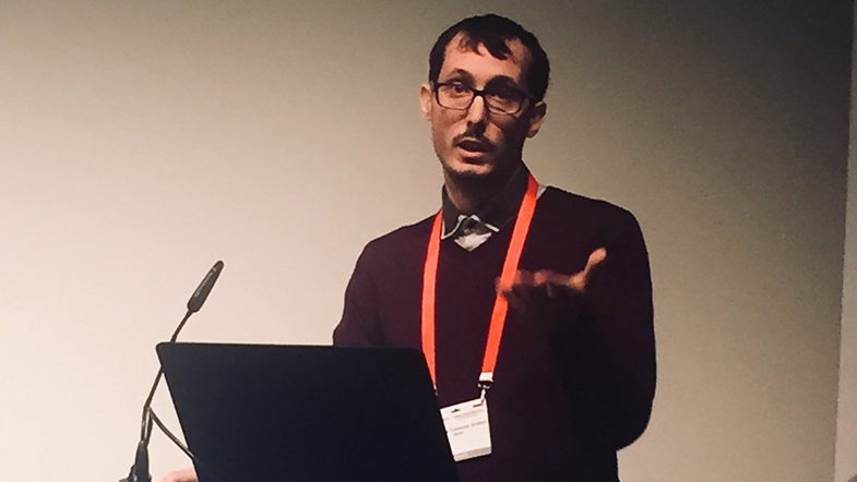
As a former Magnetic Resonance Imaging in Multiple Sclerosis (MAGNIMS) grant fellow, Mariano Cabezas participated in the MAGNIMS alumni meeting on Wednesday 10th. Mariano presented his previous research on new lesion detection developed during his stage at Vall d’Hebron Hospital in Barcelona. His resulting research has permitted clinical experts to automatically delineate new lesions on MS patients, reducing significantly the variability between experts and also the diagnosis time.


At the same time, we presented also two posters on automated lesion detection using deep learning during the conference. Sergi Valverde introduced our new pipeline nicMSlesions, which has been lately re-designed to reduce the amount of training data, allowing accurate lesion segmentation using few or even a single training image. Similarly, we also presented an extensive analysis of different state-of-the-art deep learning pipelines applied also to lesion segmentation done by our PhD student Albert Clérigues. In this work, we analyzed six different brain lesion segmentation deep learning models, studying the effect of their characteristics on the accuracy segmenting MS lesions.


The conference was an interesting opportunity to share experiences and learn from the world-top experts in the field. It was also a fantastic opportunity to have feedback from our tools and also to understand better the clinical expert needs. We strongly believe that our tools can continue contributing to the MS field, helping the clinical experts to monitor and diagnose the disease.


We participated in MICCAI 2018

MICCAI 2018, the 21st international conference on Medical Image Computing and Computer-Assisted Intervention, was held in Granada, Spain, from September 16th to 20th, 2018. The MICCAI conference attracts world-leading biomedical scientists, engineers and clinicians from a wide range of disciplines associated with medical imaging and computer-assisted intervention. The series of lectures includes three days of oral presentations and poster sessions. Besides, the day before and the day after is full of workshops, tutorials and challenges.


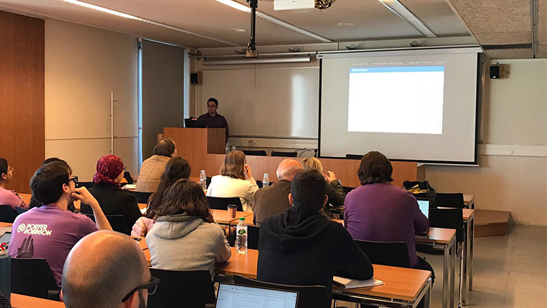
We participated in three different challenges, the MRBRAINS18, related to brain tissue segmentation, the BRATS2018, related to brain tumour segmentation and prediction, and the ISLES Challenge 2018, related to the segmentation of ischaemic stroke lesion segmentation. The responsible of presenting the works were Kaisar Kushibar, Mariano Cabezas, and Albert Clèrigues, respectively. It is worth to mention the 5th position we obtained in the ISLES challenge over more than 25 participants (we actually were the first non-Chinese participant) and the 6th position over 36 in the MRBRAINS18. Moreover, Sandra Gonzalez gave an oral lecture in the PACTH-MI workshop (Patch-Based Techniques in Medical Imaging), where she explained her proposal to extend brain multi-atlas segmentation in brains with focal lesions, like the ones of multiple sclerosis patients. This work was done in collaboration with the researchers of Vanderbilt University, where she did her research stay.


The MICCAI conference has been an excellent opportunity to talk and share experiences with top leader researchers in the world. We have seen the recent trends in medical imaging and we hope to participate also next year in Shenzhen (China).


Photo: ISLES organization

SERAM 2018: ViCOROB team represented at Biotechnology and Computing Session

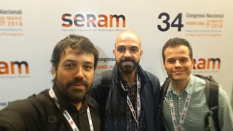
Last month, three members of VICOROB attended to the 34th National Congress of the Spanish Society of Medical Radiology SERAM and presented 3 oral and 3 poster presentations. This event represented a great opportunity to show part of the most recent research works in breast and brain imaging developed at the institute: breast parenchyma enhancement automated classification methods, image analysis tools to improve breast cancer diagnosis or deep learning approaches for multiple sclerosis’ lesion segmentation, among others.


This biennial event gathers mainly radiologists, but also other health professionals such as technicians as well as researchers and engineers with a special interest in several medical fields (breast, thorax, paediatrics, etc.). Due to the recent advances and contribution of the artificial intelligence into radiology, a new dedicated session was included this year in the congress’ program: Biotechnology and Computing, and it was in this session where VICOROB’s researchers presented their latest work. Also, during the opening ceremony, both the president of SERAM and the president of the Radiological Society of North America (RSNA) made clear that new technologies, widely used at VICOROB, such deep learning will have a great impact in the future of the radiology.


The next SERAM congress will take place in Zaragoza in 2020 and VICOROB will be there presenting their latest advances in medical imaging.


DOCTORAL THESIS: Glandular Tissue Pattern Analysis Through Multimodal MRI-Mammography Registration

By Eloy García Marcos

Supervised by Dr. Joan Martí Bonmatí / Dr. Arnau Oliver Malagelada

Abstract

Breast cancer is the most common cancer in women worldwide. Current statistics show that one in eight women will develop this disease over the course of her lifetime. While X-ray mammography is the gold standard image modality for screening and diagnosis of breast cancer, it presents decreased sensitivity in dense breasts. Several studies have shown that the combination of the different medical image modalities, such as mammography and magnetic resonance imaging (MRI), leads to a more accurate diagnosis and, therefore, a more effective medical treatment of patient diseases. However, the fusion of information among several image modalities is a challenging task, due to the differences not only in the physics underlying each modality but, also, the different patient positioning during the image acquisition. The main purpose of this thesis is to evaluate the similarity among the information provided by two medical image modalities, such as the X-ray mammography and MRI, and, at the same time, to propose new algorithms to register the images in order to correlate the position of lesion and susceptible areas.


A deep review of the state-of-the-art, focusing our attention in the multimodal registration problems using patient-specific biomechanical finite-element (FE) models, is performed, from the biomechanical model construction (including the pre-processing and segmentation of MRI images, a suitable FE mesh construction as well as the methodology to quantify the accuracy and quality of the methods) to the physics underlying the mechanical deformation (elastic and hyperelastic parameters exposed in the literature, and loading forces and boundary condition) to solve the problem.


Our analysis begins evaluating the similarity of the glandular tissue between two mammograms from the same patient, acquired in the same day and in a short time frame. This fact allows us to evaluate the effect of the breast compression in the parenchymal pattern distribution. The monomodal analysis provides us a baseline result to perform the multimodal comparison between MRI and mammography. To achieve this goal, a fully automatic framework to register the density maps, obtained from digital mammograms and MRI images, was developed. This software uses a patient-specific biomechanical model of the breast, which mimics the mammographic compression performed during the mammographic acquisition. In this work, we propose a new methodology to project the glandular tissue directly from the MRI, avoiding the loss of information that can be yielded when the image is deformed. Our analysis shows a high similarity between the information contained in the two modalities as well as a high structural similarity in the distribution of the glandular tissue.


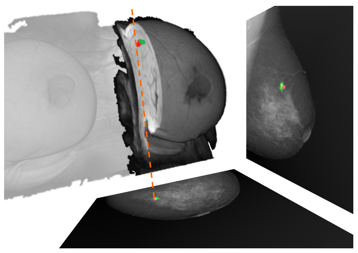
During the registration framework evaluation we computed the target registration error (TRE) between landmarks -i.e. lesions- in both MRI and mammography. The 2D problem consists of directly projecting the landmark position from the MRI to the mammogram, computing the Euclidean distance between the computed and the real landmark position. However, locating the 3D position, within the MRI, from the corresponding lesions in the mammograms, used to require complex and computational expensive methods to undo the breast compression. To solve this issue, we propose a new, fast and efficient algorithm to locate the landmark position within the MRI. Using a similar methodology to that proposed during the glandular tissue projection, a back-projection ray-tracing can be performed, allowing to locate the ray path in the uncompressed biomechanical model, avoiding to undo the breast compression. To accelerate the computation of the intersection between two rays, we propose an easy algorithm which sub-divide the rays, reducing the search space. Our model reduces the search space until 600 times with respect to a traditional point-by-point search. Thus, the computational time to locate the intersection is about 8 ms, allowing real-time applications in the clinical practice. Furthermore, the TRE, in average, are about 1 cm, better than those exposed in previous works.


To conclude, we evaluate the capability of using intensity gradient values to perform the registration between the images. In this case the glandular tissue gradient is extracted from the mammograms and MRI images by means of image processing techniques. The MRI gradient is projected to the mammogram using two different approaches. The first one uses the accumulated directional derivative, obtaining a scalar value which is comparable to the norm of the gradients obtained from the mammograms. The second one accumulates the gradient considering each direction independently, yielding a vector which is comparable to the directional derivative obtained from the mammograms by means of a gradient correlation metric. Our results show an improvement in the TRE using scalar gradient values, with respect to the traditional intensity-based approach.


In summary, this thesis will help radiologists and physicists to better understand the variations of the glandular tissue that can be clearly visible in one modality but not in the other. Furthermore, the evaluation of the information can guide researchers to obtain more accurate segmentation algorithms, considering the partial volume effect presented in the MRI, as well as to improve the multimodal image registration between the two modalities, not only by means of intensity-based methods but also considering additional information such as gradients. Our methodology includes several proposals to develop real time applications or with acceptable time values in the clinical practice.

We obtain the 4th position in two of the MICCAI 2017 conference challenges

The 14th of september, VICOROB presented two convolutional neural network methods to the iSeg and White Matter Hyperintensities challenges from the MICCAI 2017 conference.

The first challenge was aimed at segmenting brain tissues from infant brain MRI, while the second one was aimed at segmenting vascular white matter hyperintensities of adult brain MRI. The first approach was presented orally by Jose Bernal and the second one was presented orally and as a poster by Mariano Cabezas. Both approaches ranked 4th among twenty teams.


El dia 14 de setembre, el grup VICOROB va presentar dos dels seus mètodes a les competicions iSeg i White Matter Hyperintensities de la conferència MICCAI 2017.

La primera competició consistia en segmentar els teixits en imatges de ressonància magnètica del cervell d’infants, mentre que la segona consistia en segmentar lesions vasculars de matèria blanca utilitzant ressonàncies magnètiques d’adults. El primer mètode va ser presentat oralment per en Jose Bernal, mentre que el segon va ser presentat oralment i en forma de pòster per en Mariano Cabezas. Ambdós mètodes, basats en xarxes neuronals de convolucions van obtenir la quarta posició d’entre una vintena d’equips.